
Cracking the Code to the Human Face
Decades of NIDCR research are helping decipher craniofacial disorders
There’s a reason we can spot a friend in a sea of people — humans are wired to fixate on faces. We are exceptionally good at recognizing faces by teasing out subtle differences in facial features, like a square jaw, arched brows, or high cheekbones.
Did You Know?
July is National Cleft & Craniofacial Awareness & Prevention Month. Hear from NIDCR’s Clinical Director Janice Lee, M.D., D.D.S., about the work her team and others are doing to improve the lives of people with craniofacial anomalies.
For easy-to-read information on other dental, oral, and craniofacial conditions, visit our Health Info page. This information is also available in Spanish.
The uniqueness of our faces inspires poets and artists, enables facial recognition technology, and helps define our identity. Yet, what makes each of our faces one of a kind remains unclear, and what can go awry during development is even murkier.
Each year in the United States, nearly 120,000 babies are affected by birth defects, about half of which involve the face and skull, or craniofacial complex. These conditions, which range from cleft lip with or without cleft palate to premature fusion of skull plates (craniosynostosis), can impair eating, hearing, speaking, breathing, and brain development. Since the early 1960s, NIDCR-supported studies have contributed to fundamental knowledge about the genetic, molecular, cellular, and environmental factors that drive facial development. This information is fueling research that may answer why we look the way we do, how hiccups in development can lead to craniofacial anomalies, and what might be done to treat them.
“The craniofacial complex is truly unique,” said NIDCR Director Rena D’Souza, D.D.S., Ph.D. “It includes bones, soft tissues, nerves, microbial communities, immune factors, and more. What we learn from craniofacial biology is highly applicable for understanding and potentially treating a wide range of disorders affecting the entire body.”
A Head and Face Database
One way to decipher the coding of the craniofacial complex is through data — lots of it. In 2009, NIDCR launched a public repository for craniofacial data called FaceBase. The database contains biological information from humans and animals, including data on gene and protein expression, salivary gland function and development, and images of the face and skull. The goal is to provide the research community with a comprehensive database and tools to accelerate collaboration, discoveries, diagnostics, and therapeutics for craniofacial disorders.
Recent studies drawing on FaceBase data have revealed genetic regions that may drive differences in face shape in healthy humans. The data has also helped researchers identify interactions among genes and prenatal exposures to chemicals, drugs, and toxins that appear to underlie craniofacial anomalies such as cleft lip and cleft palate. These findings may one day help clinicians identify families and individuals at higher risk and offer prevention and targeted therapy.
“One of the biggest mysteries in the field is the interplay between genetics, epigenetics [reversible changes that affect gene activity], and environmental risk factors,” said Yang Chai, D.D.S., Ph.D., of the University of Southern California (USC), a co-principal investigator of the FaceBase project. “It can be difficult to tease apart the subtle contributions of multiple factors that can have nonlinear effects. We’re only beginning to unravel them.”
Tracing Development, Cell by Cell
While scientists can’t pinpoint all the causes of conditions like cleft lip and cleft palate just yet, they know certain craniofacial anomalies arise during early prenatal development. During this period, clusters of embryonic stem cells give rise to highly specialized cells that later become bone, cartilage, nerves, and soft tissue. In cleft lip and cleft palate, mishaps in this meticulously orchestrated process may disrupt the merging of the right and left sides of the lip and palate, leaving a gap, or cleft.
“We know quite a bit about the big picture of development, where cells become specialized tissues and take on specific functions,” said Lillian Shum, Ph.D., Director of NIDCR’s Division of Extramural Research. “But we know very little about how individual cells know where to go and how they got there. Imagine if we can create a craniofacial development map — we can reroute lost cells to reach their destination and prevent diseases.”
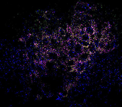
Mapping embryonic cell development is exactly what NIDCR investigator Laura Kerosuo, Ph.D., is working on. She studies the behavior of neural crest cells, which form in human embryos around the third week of development. They eventually give rise to the craniofacial skeleton, parts of the salivary glands, teeth, and more. Roughly 20% of human birth anomalies are linked to the neural crest. Understanding the normal timing and migration patterns of these cells can shed light on everything from disorders of the head and face to cancers.
Advancing Treatments: Scalpels, Algorithms, and Stem Cells
Modern medicine has made it possible to surgically treat or correct certain craniofacial conditions. Despite these advances, existing surgical techniques are often complex and highly invasive, and children may still encounter problems with feeding, breathing, and speaking after surgery. NIDCR-funded studies are looking for ways to improve surgical outcomes and better support families and patients.
Ongoing studies are optimizing surgical techniques to improve speech outcomes and facial appearance. Other scientists are working to employ advanced computer algorithms and artificial intelligence to improve evaluation of craniofacial anomalies for treatment planning. Researchers have also been assessing the impact of cleft repair on children’s oral health and social well-being, which may allow clinicians to provide more targeted care and improve quality of life.
Regenerative medicine holds promise as a less invasive way of treating craniofacial anomalies like craniosynostosis. Infants with the condition have bones in the skull that join too early, affecting head shape and brain development. Using stem cells to regenerate the tissue that separates skull joints, a USC team led by Dr. Chai corrected skull shape and reversed learning and memory deficits in young mice with craniosynostosis. The research opens the door to stem cell therapy as a more effective, less invasive means of treating craniosynostosis and similar disorders in humans.
Bringing Discoveries to Life
“The time has come to coalesce various knowledge fields so that surgical interventions are not the only options available to patients affected by inherited disorders of the craniofacial complex,” said Dr. D’Souza. “This is one of the most promising outcomes of FaceBase — this unique data-sharing platform guides clinical decision making in a manner that improves early diagnosis, screening, and treatment protocols for our patients.”
Looking forward into the next decade, scientists foresee big data and advanced computer algorithms such as artificial intelligence propelling precision medicine into the spotlight. These scientific strides will reveal the secrets behind our faces and drive tailored prevention, diagnosis, and treatment for dental, oral, and craniofacial disorders.
“The amount of knowledge and data we are collecting is breathtaking,” said Dr. Shum. “With these data, we are poised to bring discoveries, answers, and solutions to the patients.”
Related Links
References
Brinkley JF, Fisher S, Harris MP, Holmes G, Hooper JE, Jabs EW, et, al. The FaceBase Consortium: a comprehensive resource for craniofacial researchers. Development. 2016 Jul 15;143(14):2677-88. doi: 10.1242/dev.135434. Epub 2016 Jun 10.
Yu M, Ma L, Yuan Y, Ye X, Montagne A, He J, et al. Cranial Suture Regeneration Mitigates Skull and Neurocognitive Defects in Craniosynostosis. Cell. 2021 Jan 7;184(1):243-256.e18. doi: 10.1016/j.cell.2020.11.037.
Attention Editors
Reprint this article in your own publication or post to your website. NIDCR News articles are not copyrighted. Please acknowledge NIH's National Institute of Dental and Craniofacial Research as the source.
July 2024