
NIDCR Digital Library
The NIDCR Digital Library provides images that are free to use with credit. Images are meant for use by the science and health community, the press that covers health and science, teachers and other educators in health and science, and non-profit organizations that produce health and science information. It is not intended for commercial use.
Media ID#: 22741
Media ID#: 22741
Developmental markers in healthy skeleton
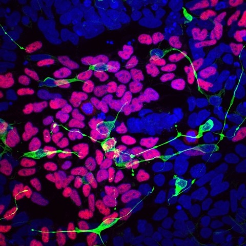
Researchers have discovered a new genetic disorder named LINKED. Compared to a disease-free individual (shown above), differentiated LINKED patient cells lack markers of normal development of the brain, spinal cord and craniofacial skeleton (pink, green, & yellow).
Media ID#: 22746
Media ID#: 22746
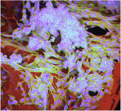
Salivary gland diversity starts early
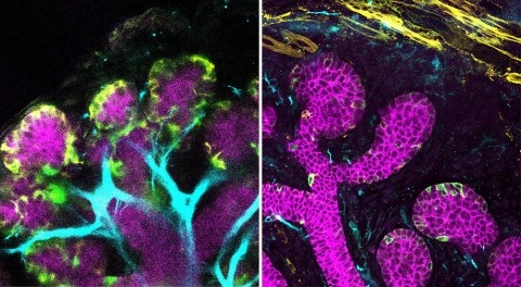
NIDCR scientists Rei Sekiguchi, DDS, PhD, Ken Yamada, PhD, and colleagues found striking differences in muscle- and nerve-related gene activity, or expression, in embryonic mouse submandibular glands (above left) and parotid glands (above right).
Media ID#: 22751
Media ID#: 22751
Exploring AI for cancer diagnosis
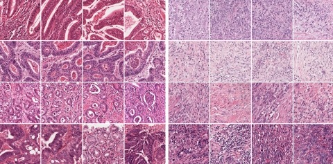
By detecting different visual patterns in tumor images, the deep learning program accurately predicted the presence or absence of molecular alterations, such as the mutated AMER1 gene, which is present in the gastric tumor images on the left but not the right.
Media ID#: 22756
Media ID#: 22756
3D salivary spheroids release amylase upon stimulation
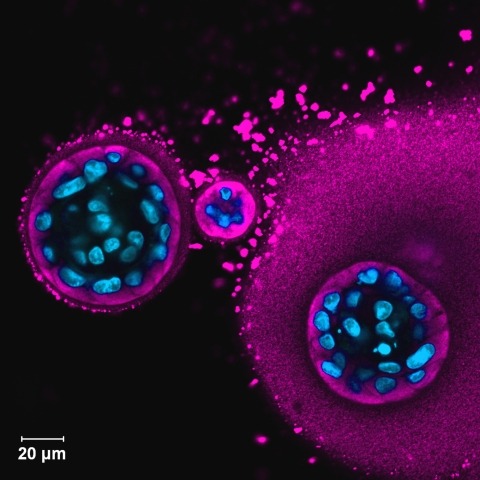
Salivary gland spheroids in hydrogels release amylase, an enzyme found in saliva, upon stimulation with neurotransmitters agonists.
Media ID#: 22761
Media ID#: 22761
Injectable tumor cell infused anti-cancer cryogel vaccine
Cancerous melanoma cells, shown with their cell bodies (green) and nuclei (blue), are nestled in tiny hollow lumens (tubes) within the cryogel (red) structure.
Media ID#: 22766
Media ID#: 22766
Stem cells in 3D hydrogel
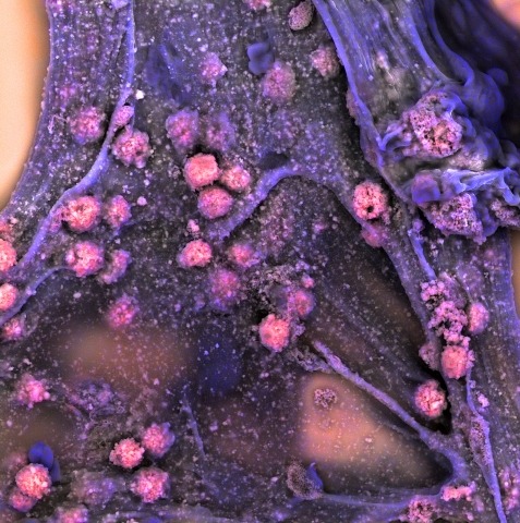
Scanning electron micrograph of mesenchymal stem cells cultured in an alginate hydrogel. The colors show the elemental composition of the sample: magenta represents phosphorus from the minerals deposited by the differentiated cells, and blue represents carbon from the hydrogel.
Media ID#: 22771
Media ID#: 22771
Embryonic submandibular salivary gland
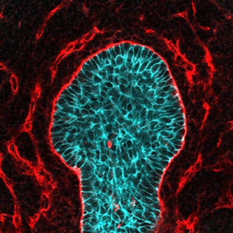
A single-bud submandibular salivary gland from a mouse embryo. The image shows epithelial cell-cell junctions (blue-green) and O-glycosylated proteins (red).
Media ID#: 22776
Media ID#: 22776
Blind cavefish neuromasts
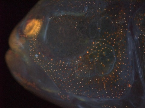
Cave-dwelling fish demonstrate frequent fragmentation of facial dermal bones. One bone is labeled beneath the eye (light blue). Cellular interactions between sensory neuromasts (labeled as orange “dots”) and the bone primordia are providing clues for scientists to understand why.
Media ID#: 22781
Media ID#: 22781
Peekaboo
A face appears when basement membrane patterns collagen IV (green) are studied in human parotid tissue where cell nuclei (white) and filamentous actin (blue) highlight the ducts and blood vessels.
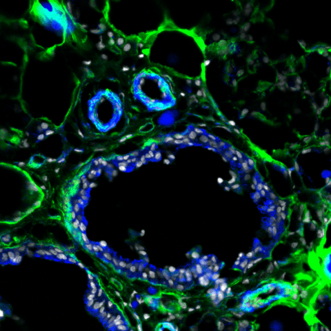
Media ID#: 22786
Media ID#: 22786
It's a small world
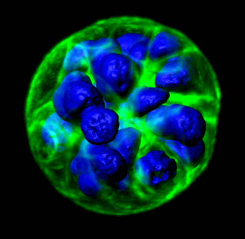
hS/PCs form three-dimensional multicellular structures (nuclei in blue) that dynamically organize and mature into coordinated units that respond to neurotransmitters (filamentous actin in green). Organization of these cells into functional secretory units is a critical for dry mouth.