
NIDCR Digital Library
The NIDCR Digital Library provides images that are free to use with credit. Images are meant for use by the science and health community, the press that covers health and science, teachers and other educators in health and science, and non-profit organizations that produce health and science information. It is not intended for commercial use.
Media ID#: 22961
Media ID#: 22961
3D Permeation
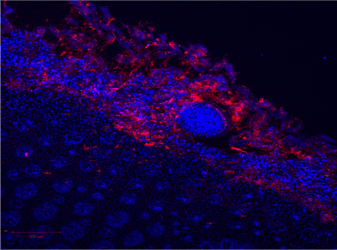
A 3D confocal image of nano-particles permeating topically into the tongue tissue.
Media ID#: 22966
Media ID#: 22966
A close look at a fruit fly salivary gland
This up-close look at a fruit fly salivary gland shows individual cells containing many secretory granules (blue-green), which store proteins that aid in the fly’s lifecycle. The black region in the middle of each cell is the nucleus.
Media ID#: 22971
Media ID#: 22971
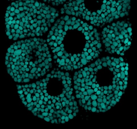
Full fruit fly salivary gland
Cells in fruit fly salivary glands are filled with cellular packets known as secretory granules (blue-green), which store molecules that aid in the fly lifecycle.
Media ID#: 22976
Media ID#: 22976
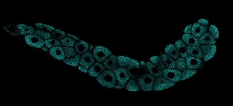
Fruit fly digestive system

This image shows part of a digestive organ called a proventriculus from a fruit fly larva. Cells in the organ’s outer layer are active in secreting a lining that protects the whole digestive system, similar to the mucous lining of the human digestive system.
Media ID#: 22981
Media ID#: 22981
Creatures-crawling-within.mp4
NIDCR scientists used live-cell imaging to capture fibroblasts (pink) using a “front wheel drive” method to propel themselves forward through a web of proteins (green) in a lab dish meant to mimic its 3-D environment in the human body.
Media ID#: 22986
Media ID#: 22986
Fruit fly salivary gland
Understanding fruit fly glands and the cellular packets known as secretory granules (red), which store proteins destined for secretion, may help scientists better understand how secretion goes wrong, and how it might be treated, in conditions like salivary gland disorders.
Media ID#: 22991
Media ID#: 22991
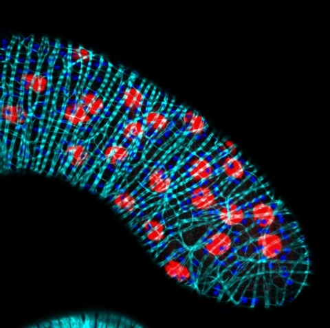
Cross section of mouse embryo torso
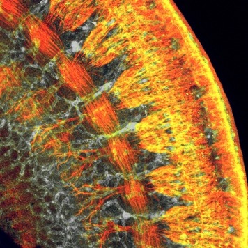
Confocal microscopy was used to create this image of a mouse embryo torso which sheds new light on mammalian development.
Media ID#: 23001
Media ID#: 23001
Salivary gland cells infected with SARS-CoV-2
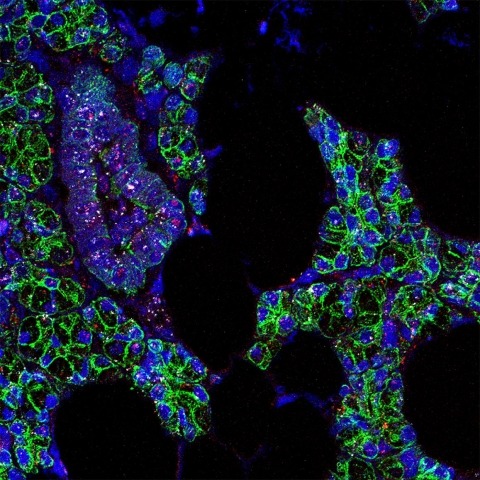
An international team of scientists has found evidence that SARS-CoV-2, the virus that causes COVID-19, infects cells in the mouth. While it’s well known that the upper airways and lungs are primary sites of SARS-CoV-2 infection, there are clues the virus can infect cells in other parts of the body.
Media ID#: 23006
Media ID#: 23006
An itch to scratch
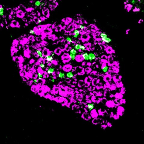
Nerves that stimulate skin are grouped in structures next to the spinal cord. Here, nerves in such a structure—called a dorsal root ganglion—that are involved in detecting an itch are labeled green. Nerves involved in sensing pain, temperature and other stimuli are shown in magenta.
Media ID#: 23181
Media ID#: 23181
Creepy crawlies on the teeth
This creeping creature is composed of cavity-causing bacteria (green) that piggyback on fungi (blue), forming superorganisms that “walk” and “lunge” on tooth-like surfaces. These movements allow the microbial assemblages to spread faster and farther, making them extra skilled at promoting tooth decay.