
NIDCR Digital Library
The NIDCR Digital Library provides images that are free to use with credit. Images are meant for use by the science and health community, the press that covers health and science, teachers and other educators in health and science, and non-profit organizations that produce health and science information. It is not intended for commercial use.
Media ID#: 22451
Media ID#: 22451
Bacterial-fungal clusters in saliva
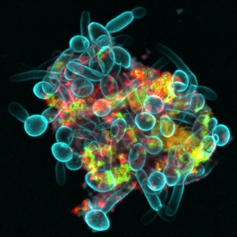
An interkingdom assemblage formed by fungi (Candida albicans in blue), bacteria (Streptococcus mutans in green), and bacteria-derived extracellular polymers (α-glucans in red) in human saliva.
Media ID#: 22691
Media ID#: 22691
Embryonic smooth muscle cell
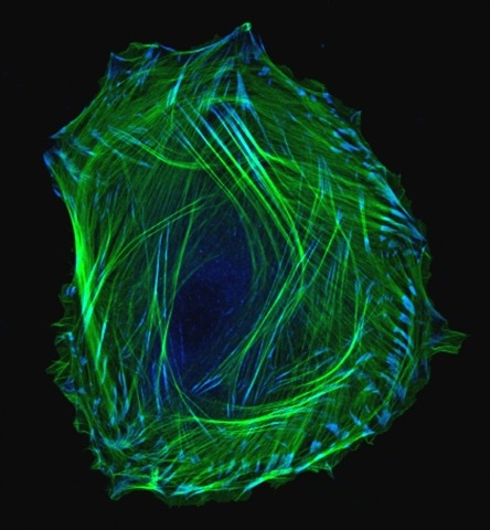
Embryonic smooth muscle cell. Immuno-fluorescently labeled actin cytoskeleton (green) and vinculin in cell adhesions (blue). Laser scanning confocal microscopy.
Media ID#: 22696
Media ID#: 22696
3D microCT E18.5 mouse skull
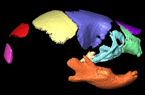
Three dimensional (3D) micro-computed tomography (microCT) analysis is used to examine the phenotypes of craniofacial bones in different mouse models
Media ID#: 22701
Media ID#: 22701
Mesoporous silica microparticles
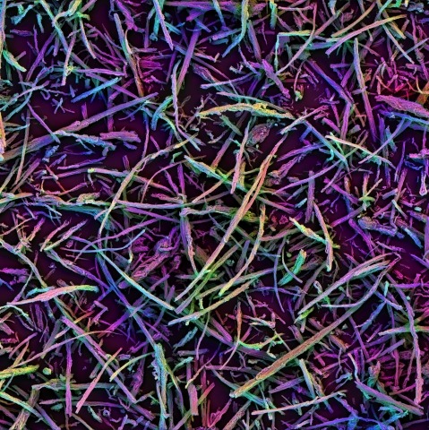
Mesoporous silica microparticles that spontaneously assemble in vivo to form 3D scaffold that allows for in situ immune cell recruitment and programming.
Media ID#: 22706
Media ID#: 22706
Lip print patterns
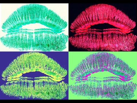
Physical features such as lip print patterns develop early in prenatal life, are unique and unchanged throughout life.
Media ID#: 22711
Media ID#: 22711
Cross sections through the orbicularis oris muscle
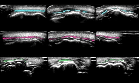
These images are cross sections of the upper lip taken by high resolution ultrasound in order to visualize the orbicularis oris muscles—colored in the images.
Media ID#: 22716
Media ID#: 22716
A cancerous conversation fuels oral tumors
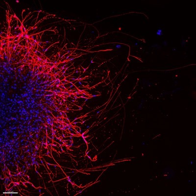
Oral cancer cells send growth signals to nearby mouse sensory neurons, which sprout projections called neurites (red).
Media ID#: 22721
Media ID#: 22721
Scientists chew on a new theory of swallowing
Media ID#: 22726
Media ID#: 22726
Immune cells patrol the brain
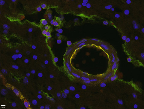
In human brain tissue, NIDCR researcher Eva Mezey and colleagues found lymphatic vessel cells (green) lining the space surrounding blood vessels (circular structure), as well as T cells (red), as shown in this cross-section.
Media ID#: 22731
Media ID#: 22731
Mimicking mother nature to grow an artificial gland

Using a technique called two-photon microscopy, researchers tracked the movements of embryonic salivary gland cells in real time. The process, budding, occurs many times to drive a gland’s growth into a mature organ that contains thousands of saliva-secreting, globe-like structures.