
NIDCR Grand Rounds lecture, Feb 2019
For anyone who dreads going to the dentist, there may be light at the end of the tunnel. Advances in laser and light-based imaging technologies may soon change the face of modern dentistry.
Tooth enamel is almost transparent at longer wavelengths, making it possible to shine near-infrared light on a tooth to detect dental decay.
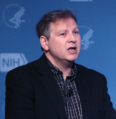
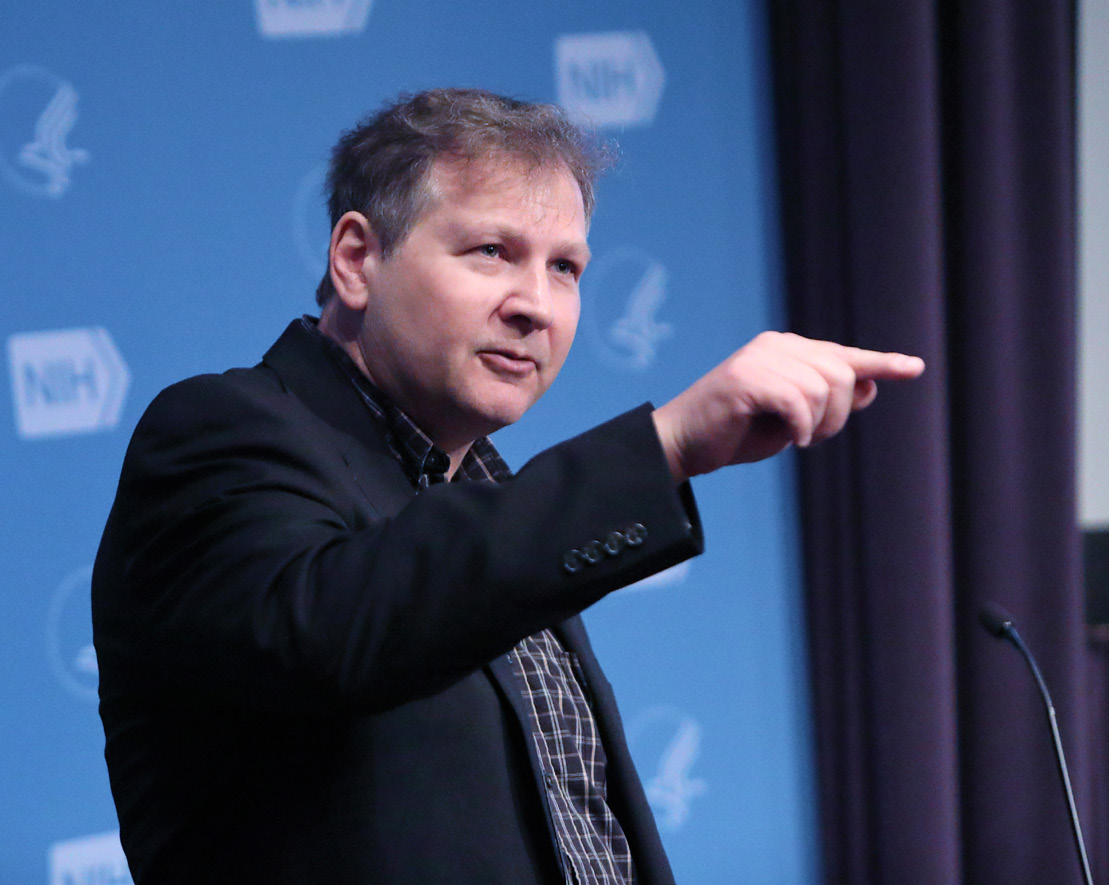
“You can see right into the tooth,” said Dr. Daniel Fried, professor, University of California, San Francisco School of Dentistry. “The enamel looks almost like an ice cube.”
Light-based imaging is minimally invasive, providing a safer alternative to an X-ray’s ionizing radiation, explained Fried at a recent NIDCR Grand Rounds in Lipsett Amphitheater. His team is also researching laser technology that can remove dental decay and composite fillings, bonding and adhesives, which could mean less painful visits to the dentist.
There’s long been a need for more reliable methods to diagnose tooth decay, said Fried. Most cavities form on the occlusal surfaces of teeth. Dentists visually inspect teeth for decay, which can lead to false-positives and overtreatment. Even X-rays are not sensitive enough to detect early occlusal cavities.
“Many lesions in the mouth have been re-mineralized and...no longer need inter¬vention,” said Fried. “Dentists have trouble telling the difference between active and arrested lesions; this new technology has the potential of differentiating them.”
Light-Based Imaging More Precise
Fried’s research focuses on two kinds of light-based imaging that provide a more precise picture than X-rays and therefore could help diagnose and treat tooth decay much earlier. Near-infrared imaging is sensitive enough to detect early demineralization and can screen many teeth at once. Optical coherence tomography (OCT), similar to an ultrasound, shows cross-sections and can image deep into the tooth.
“OCT [already] has changed the practice of ophthalmology,” said Fried. “It’s been very successful for
retinal imaging...and it’s also very promising for dentistry.”
Capable of imaging through composites and sealants, OCT is particularly useful for assessing lesion severity and activity. “If the dentist doesn’t know if [a lesion] is active or arrested,” said Fried, “with OCT, you can actually see the lesion structure, how deep it is and if it has a definitive surface zone suggesting that remineralization has occurred.”
Tomography is especially suited for clinical trials as it can track changes over time. In OCT clinical trials, Fried’s lab has detected significant demineralization that wasn’t spotted visibly. He recalled that their first studies in 2010 were encumbered by slow technology. Now, they’ve acquired a new system that uses a scanning device on a chip capable of taking entire 3-D images in a second.
“One of the most exciting things we can do with OCT is monitor the changes in lesions as we re-mineralize them,” said Fried. “With nonsurgical intervention, you can treat [the tooth] with fluoride and re-mineralize lesions...That’s important for assessing lesion activity” and whether intervention is necessary.
In a demineralized tooth, the decay reflects a lot of light and appears white against the healthy enamel, which looks dark in the near-infrared.
“We get the highest contrast at these longer wavelengths, significantly higher than other imaging technologies,” said Fried. And there’s another benefit to near-infrared imaging. “Stains, which are responsible for a lot of false-positives, don’t absorb at these longer wavelengths, so you can image just the demineralization without the stain.”
A recent clinical study found a dramatic difference between near-infrared imaging and X-rays, reported Fried. In 26 lesions seen at the near-infrared that penetrated the dentin, only 1 of them showed up on X-ray.
Lasers Can Remove Cavities
You may know the drill. Now meet the new lasers that can selectively remove cavities. Compact and precise, these infrared lasers scan the tooth’s surface and emit tiny, fast pulses to remove decay selectively without overly impacting healthy tooth structure.
Fried’s research focuses on two kinds of light-based imaging that provide a more precise picture than X-rays and therefore could help diagnose and treat tooth decay much earlier.
The three technologies complement each other. First, a near-infrared image is taken and Fried’s team
has an algorithm to convert it to pixels. Then the high-speed laser scans the surface and removes decay, followed by an OCT scan that checks how much was removed.
“You can also use the near-infrared to enhance visibility of composites,” said Fried. “Dentists spend more time removing existing composites and restorations than putting in new ones. If the composite is color-matched to the tooth, it’s hard to see where it is,” and that makes it tough to remove without damaging nearby healthy enamel.
Spectral-guided ablation can be used to selectively remove composite from tooth surfaces. When a laser strikes material, some of it vaporizes and the plume looks different when it strikes enamel vs. composite, explained Fried. Using spectral-guided ablation, it’s possible to see the calcium lines of enamel to make sure the laser is only striking composite. In a current clinical study, Fried’s lab is removing small composite restorations in less than a minute.
The combination of these laser and light-based technologies could lead to earlier and better detection and intervention of dental decay. So smile! The future of dentistry is looking bright.
–By Dana Talesnik
Related Links
• Novel Light-Based Technologies for the Detection, Diagnosis, & Selective Removal of Dental Decay
• Dental Materials and Biomaterials Program
• Tooth Decay
• Fillings
References:
Chan KH, Fried D. Multispectral cross-polarization reflectance measurements suggest high contrast of demineralization on tooth surfaces at wavelengths beyond 1300 nm due to reduced light scattering in sound enamel. J Biomed Opt. 2018 Jun;23(6):1-4. doi: 10.1117/1.JBO.23.6.060501.
Yang, V. B, Curtis, D. A., Fried, D. Use of Optical Clearing Agents for Imaging Root Surfaces with Optical Coherence Tomography. J. Sel. Topics Quant. Elect. 25(1), 1-7 (2018).
Funding: NIH’s National Institute of Dental and Craniofacial Research (NIDCR)
Adapted from an article in the NIH Record, February 22, 2019.