Scientific Application of Advanced Imaging Technology
Facilitating the research goals of our Intramural research community through the application of cutting-edge microscopy approaches.
Mission
The NIDCR Imaging Core is a centralized facility that provides imaging resources to the NIDCR intramural research community. We provide an expert knowledge base and maintain numerous state-of-the-art imaging platforms in support of our researchers. Support from Core staff covers the entire lifecycle of an experiment and can involve assistance with experimental design, sample preparation, image acquisition, and data analysis. Open access to our microscopes is provided following an initial consultation and training on the relevant microscope.
Capabilities
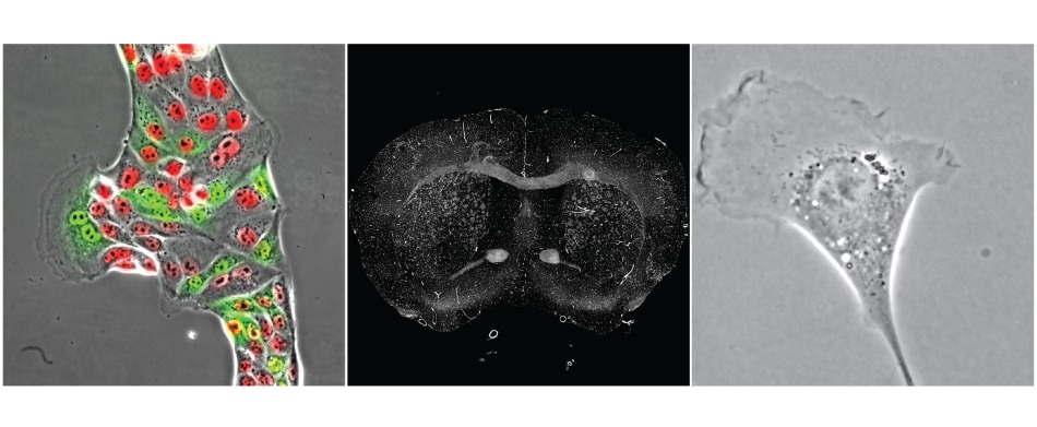
Automated Widefield Imaging
Microscopes are available for camera-based acquisition of brightfield and multi-color fluorescent imaging. Each system is equipped with an environmental chamber that provides heat, humidity, and CO2 for live imaging applications. Imaging software provides the ability to automate acquisition of high temporal and spatial imaging data.
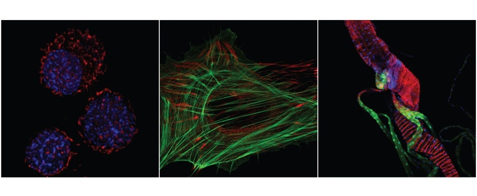
Confocal Based Imaging
Microscopes are available for a vast array of confocal based imaging applications using both fixed and living samples. Some of our capabilities include: high resolution multi-color imaging, high speed multi-color real-time imaging, deep tissue multiphoton imaging, spectral imaging, FRAP and FRET.
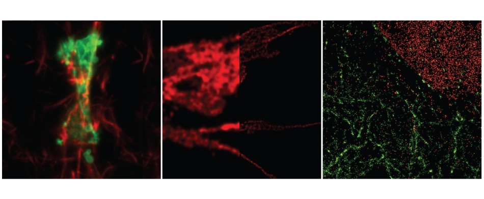
Super Resolution Imaging
Microscopes are available for various imaging applications that require resolutions greater than what can be attained on a conventional diffraction limited system. These microscopes cover a broad range of super resolution modalities including STORM, STED, SIM, array detector, and isotropic light sheet microscopy.