
Neural Crest Development & Disease Unit: Research
Research
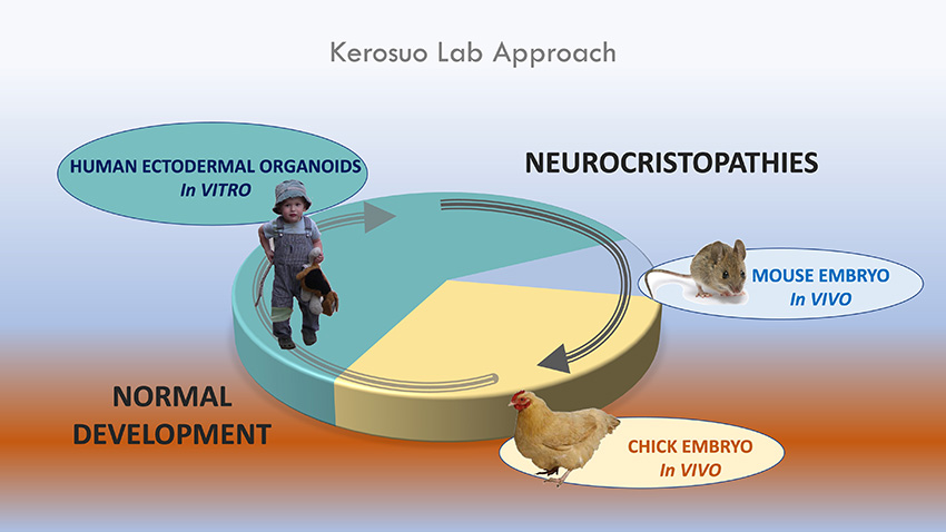
The overall aim of the Kerosuo lab is to provide a comprehensive picture of early neural crest development as part of the ectoderm patterning process and neurulation. In addition to gaining information on normal development, we use this knowledge to unravel the pathology behind neural crest-derived diseases known as neurocristopathies, which include roughly a quarter of birthdefects, such as lip and cleft palate, Hirsprung’s disease, CHARGE, DiGeorge, and Treacher Collins syndromes and cancers like melanoma and neuroblastoma. We focus on understanding the molecular mechanisms behind neural crest pluripotency-like stem cell maintenance, how fate choices are made, and the extent of heterogeneity and plasticity in neural crest potential. To answer our research questions, we use a combination of cutting-edge cell and molecular biology techniques and single cell and live imaging on chick and mouse embryos as well as on human pluripotent-cell-derived neural crest cells. By combining the induced pluripotent stem cell (iPSC)-technology to our research, the goal is to create a bridge between normal development and disease and create neural crest cells from patients with neurocristopathies to characterize the underlying molecular cause, which in part, are further validated by using the in vivo animal models.
Our Focus
Neural crest cells consist of a population of pluripotent-like stem cells found in early vertebrate embryos. They give rise to more than thirty different cell types and tissues ranging from ganglia of the peripheral nervous system, bone and cartilage of the face, cornea in the eye, pigmentation of the whole body, and multiple types of endocrine cells. The repertoire is exceptionally broad and diverse given the fact that the neural crest is derived from the post-gastrulation ectodermal germ layer that already has taken primary steps towards fate commitment.
A major goal of the Kerosuo lab is to understand how neural crest cells acquire and regulate their exceptionally high stem cell potential in the ectoderm and subsequently control their commitment into progenitors and various differentiated cell types during early embryo development. We use a broad range of modern era molecular biology approaches including high resolution imaging, single-cell RNA sequencing (scRNASeq), mass spectrometry, and single-cell Multiplex Spatial Transcriptomics (scMST) technique to reach our goal. Our second major focus area is to connect normal embryo development to disease. Dissecting the molecular details during the normal developmental process will lead us to understanding how neural crest-derived cancers and birth defects (a.k.a. neurocristopathies) are formed.
Recent Findings in Neural Crest Development
Our recent work (Pajanoja, et al., Nature Communications, 2023) has shown that the entire ectoderm, and not just the neural crest domain, maintains a pluripotency-like signature after gastrulation, which challenges the dogmatic viewpoints of permanent loss of pluripotency during gastrulation. Our findings also propose that this maintenance of the pluripotency signature from the blastula stage is the mechanism by which the neural crest gains its exceptionally high stem cell potential and provides much higher plasticity during the ectodermal patterning process than previously thought.
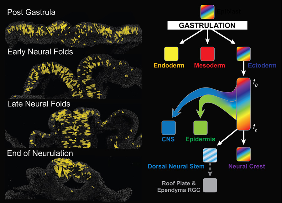
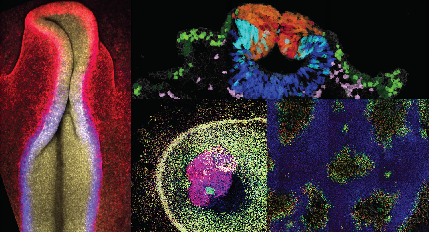
Left: Cross sections from a neurulating chick head shows how cells with a pluripotency signature are not lost during gastrulation but instead are found throughout the entire ectoderm and are then gradually restricted to the dorsal neural tube at the end of neurulation. The pluripotent-like cells are pseudo-colored yellow in the original images by using our custom-developed single cell Multiplex Spatial Transcriptomics technique. Right: We hypothesize that expression of pluripotency genes (Nanog, oct4, Llf4, etc.) continue to be expressed in the entire ectoderm after gastrulation, which ensures a gradual ectodermal patterning process to the different domains. While the pluripotency is gradually lost in the future CNS and epidermal domains, respectively, during mid-neurula stage, it is maintained in the dorsal neural tube that forms the neural crest and the dorsal neural stem cells that give rise to the roof plate. We hypothesize these are the mechanisms by which the neural crest gains its exceptionally high, pluripotency-like stem cell potential. (Images adopted from Pajanoja et al., Nature Communications, 2023).
Development of Tools to Make Discoveries
The usage of our spatial transcriptomics method (scMST, which is an optimized version of our previous single-cell Multiplex Spatial Transcriptomics technique to address transcriptional profiles in multiple imaging fields from multiple developmental stages) was key to our discovery of the pluripotency signature maintenance in the developing ectoderm. With this technique one can determine transcriptionally distinct subpopulations of cells from a heatmap and visualize them on a single cell resolution within the original 3D tissue by pseudo-coloring the cells from different subpopulations. This allowed us to detect intermingling cells with different stem cell states from all ectodermal domains to build our hypothesis (Pajanoja, et al., Nature Communications, 2023).
We have also developed ShapeMetrics, a user-friendly pipeline for 3D cell segmentation and spatial tissue analysis on microscope images, which provides single cell level measurements of cell sizes and shapes within the original tissue. Please visit our GitHub page to access the codes.
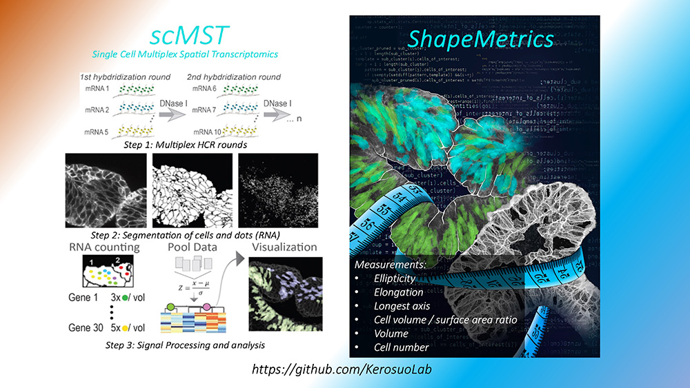
The Neural Crest Development & Disease Unit has invested in developing tools to accurately answer its research questions. scMST is based on re-hybridization rounds of single molecule fluorescent in situ hybridization. Individual cells are segmented in 3D from spinning disk confocal images, and the transcripts are counted within each cell. The single cell transcription data is pooled into a heatmap; transcriptionally distinct subpopulations are assigned a color, and the pseudo-colored cells are visualized in the original cross-sections to show the spatial locations of the subpopulations. ShapeMetrics is based on the same 3D cell segmentation as scMST. The shape of the individual cells is measured, and cells can be separated to subgroups in a heatmap based on their morphology features and pseudo-colored to visualize the location in the original images. Please visit our GitHub page to access the codes.
Current Research Questions
Our recent discoveries raise new questions that we are currently investigating: What are the molecular mechanisms behind the neural crest pluripotency-like signature regulation? During what stage of neural crest development do we start detecting signs of lineage commitment and respective subpopulations? How does the ectoderm get patterned? Is the neural crest stem cell population the same ‘from head to toe’, throughout all axial levels from anterior to posterior? Can we accurately model human neural crest by using 3D in vitro approaches? Which neurocristopathies are caused by defects in neural crest stem cell regulation? What can we learn about neuroblastoma initiation by comparisons with normal neural crest? To what extent is DiGeorge syndrome a neurocristopathy?
Embryo Work and Human Disease Modeling
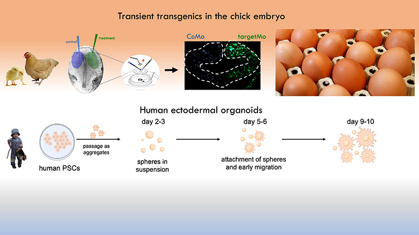
To answer our research questions, we use an array of cutting-edge molecular biology techniques both in the chicken embryo in vivo, as well as by using human embryonic stem (ES) cell-derived neural crest cell cultures. The advantage of the chicken embryo technique is its amenability to tissue-targeted transient transgenics using in ovo and ex ovo electroporation. As amniotes, early chicken embryo development is highly similar to humans. Yet, due to their development outside the mother, chicken embryos allow detailed investigation and manipulation at early developmental stages, which often is challenging in mammalian model systems. To study late developmental phenotypes, we use the mouse model. In addition, the usage of human ES and iPSC-derived neural crest cells as our in vitro system allows us to perform molecular and functional studies on human cells. This approach also provides the opportunity to model and study disease mechanisms in patient-derived induced neural crest cells.
Principal Investigator

November 2024