
Craniofacial Anomalies & Regeneration Section: Learn About Our Research
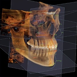
Human and Mouse Craniofacial Anomalies
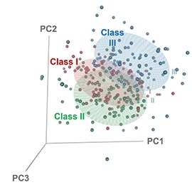
Anomalies found in human disorders provide an important window to both normal and abnormal development. The mission of the Lee lab is to understand craniofacial and dental development by studying human disorders that affect normal development and growth of the craniofacial skeleton, soft tissue, and dentition, develop prediction modeling of craniofacial development in healthy and disease cohorts and identify non-invasive target therapies to correct these deformities and anomalies. Our approach includes a combination of clinical, translational, and basic research with the use of state-of-the-art imaging and deep quantitative analysis techniques.
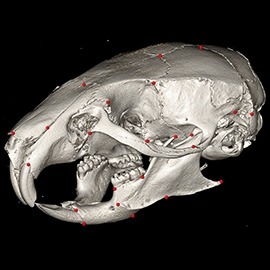
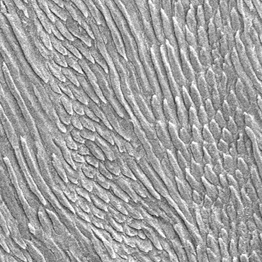
Human and Mouse Dental and Enamel Anomalies
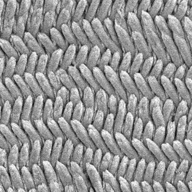
Some of the patients with craniofacial anomalies also exhibit dental anomalies. Our lab is interested in thoroughly characterizing these dental defects that result in teeth fractures and abnormal shape. This includes the ultrastructural and biomechanical analysis of dental specimens from patients as well as the use of animal models that recapitulate the phenotype of the disease. These animal models are used to further understand the developmental alterations that lead to the phenotype and the molecular mechanism underlying these alterations.
Bone Regeneration
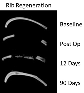
Aging negatively impacts human bone regeneration capacity. The longstanding interest of our lab is to characterize age-related changes at the molecular, cellular, and functional levels in large-scale bone defects that represent a challenge in clinical practice. For this purpose, we are using a rib resection mouse model that recapitulates the process of bone regeneration in humans. Our ultimate goal is to develop new effective therapeutics to enhance bone repair and the functional performance of large bone defects.
April 2026